How to Prepare for an Echocardiogram

Blue Daily
| 4 min read

Echocardiograms are a common, non-invasive diagnostic test that shows pictures of the heart and its valves. This test is useful to look for congenital heart disease, review damage from heart attacks or heart failure and to check for problems with the inner workings of the heart. They can be requested by your primary care provider or a cardiologist to examine the workings of the heart and its blood vessels. The test will often be done at a hospital or clinic.
What is an echocardiogram?
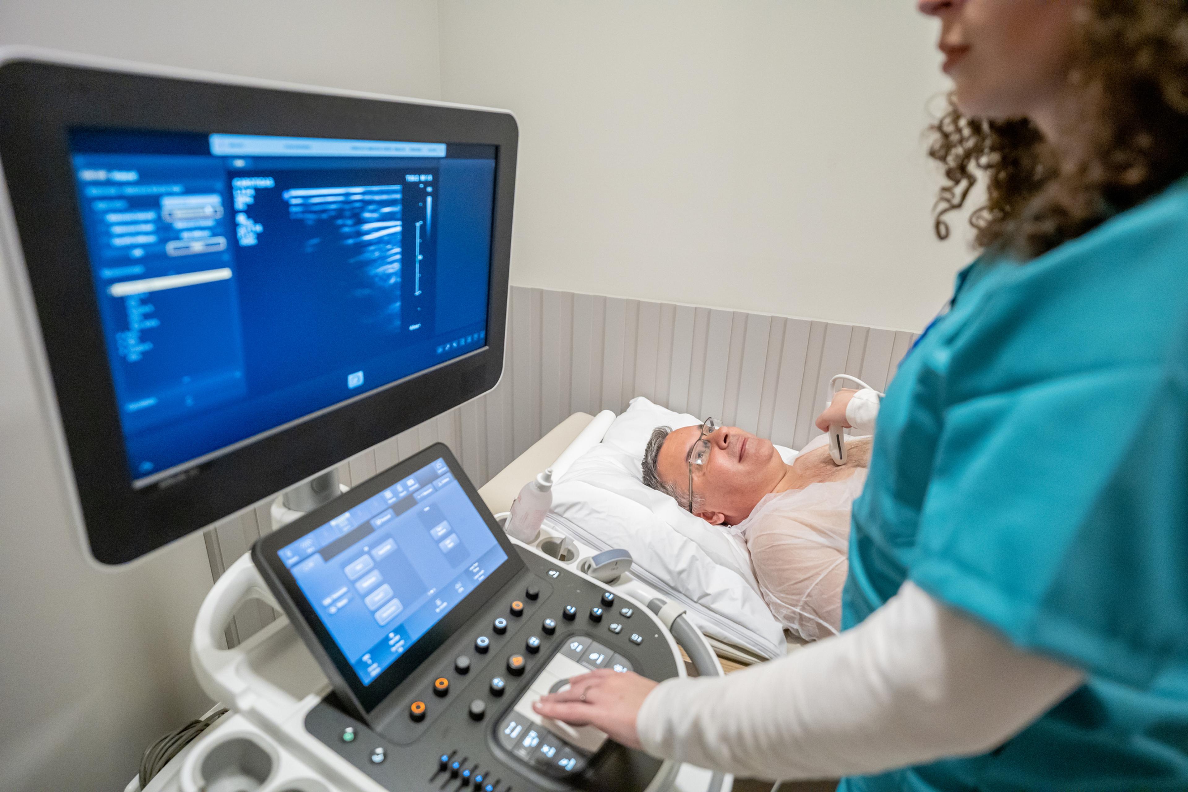
An echocardiogram is an ultrasound of the heart muscle and valves conducted by a cardiac sonographer or echocardiographer. The ultrasound uses sound waves to make moving pictures of the heart. There are several different types of echocardiograms.
Health care providers often call these “echos” for short.
Why would I need an echocardiogram?
Doctors often order echocardiograms to look for signs of heart disease, track the progression of heart disease and check in to see how well medical and surgical treatments are working.
What do echocardiograms show?
Echocardiograms can give the health care team a better view and understanding of the inner workings of the heart. They can show any problems with heart valves, heart defects you were born with, blood clots, tumors, muscle weaknesses and any enlarged portions of the heart or thickness in the lower chambers. Echocardiograms can be used to diagnose heart problems.
How long do echocardiograms last?
The length of the test depends on the type of echocardiogram that is being done. They can last from 10 minutes to two hours.
Types of echocardiograms
There are several different types of echocardiograms:
- Transthoracic echocardiogram (TTE): This is the standard echo test. Electrodes are placed on your chest, and the technician will run a gel-covered wand over several areas of your chest to generate images. Contrast dye may be given by IV to improve the quality of the images.
- Transesophageal cardiogram (TEE): This test requires an ultrasound probe to be placed into the throat and down the esophagus. The location is close to the heart, allowing a clearer picture. Often, this is performed under mild sedation.
- Stress echocardiogram: This is performed while you are exercising on a treadmill or stationary bicycle. You first undergo a transthoracic echocardiogram, then you will exercise, and then you will undergo another transthoracic echocardiogram.
- Fetal echocardiogram: This is used to take pictures of a baby’s heart while they are still in the womb. It can be used to evaluate if the baby is at risk for congenital heart defects. These are performed either through an abdominal ultrasound or through an endovaginal ultrasound.
How is this different from an EKG?
An EKG, or electrocardiogram, is another common test of the heart’s functionality. It uses electrodes to check how the heart is working, and is a good first line test to show if the heart is beating normally or irregularly. It can help detect patterns, and indicate if there are issues with the heart’s shape or size that need to be tested further.
However, an EKG is not effective in evaluating if the heart is pumping well.
An echocardiogram is able to show health care providers more details about how the heart valves are working, including how the ventricles are communicating, the speed at which blood leaves the heart and additional information about the size and shape of the heart.
How to prepare for an echocardiogram
Ask your health care provider for specific instructions about how to prepare for your echocardiogram. There are different types of echocardiograms that may require different types of preparations.
For example, some echocardiograms will require you to take off your clothes from the waist up and put on a hospital gown. Additionally, you may be asked to not eat or drink anything for a certain amount of time before the test. Avoiding caffeine, smoking and certain medications may be another requirement.
Follow your health care provider’s instructions to prepare for the echocardiogram to ensure the test goes smoothly.


